
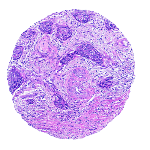
Cervical Tumour Tissue Microarray for Cancer Research
Tissue microarrays (TMAs) are an advanced research tool that allows the simultaneous analysis of multiple tissue samples on a single slide. By embedding standardized cores from different donor tissues into one paraffin block, TMAs make it possible to conduct high-throughput studies with reduced variability and cost. A cervical tumour tissue microarray supports cancer research by enabling the examination of key biomarkers related to cervical carcinogenesis, such as HPV-related protein expression, tumour progression patterns, and patient-specific variations across a diverse cohort.
Cervical tumour TMAs are primarily used in immunohistochemistry (IHC) and hematoxylin and eosin (H&E) staining. IHC is commonly applied to study the expression of viral oncoproteins like HPV E6/E7, p16INK4a, and other molecular targets linked to cervical cancer diagnosis and prognosis. H&E staining provides a detailed view of tissue structure, allowing researchers to evaluate tumour morphology, epithelial changes, and histological grading. Together, these methods help researchers investigate the molecular pathways of cervical cancer and validate potential biomarkers for early detection and treatment.
Role of Tissue Microarrays in TME Research
Tissue microarrays have transformed how researchers explore the tumour microenvironment (TME). They enable high-throughput analysis of multiple tissue samples simultaneously, making them essential for identifying biomarkers, conducting immune profiling, and performing spatial analysis. TMAs allow researchers to map immune cell infiltration, tumour heterogeneity, and stromal interactions across a broad cohort. Because human-derived TMAs preserve spatial organization, they provide a more accurate representation of the TME compared to animal models. This accelerates the validation of predictive biomarkers and improves the understanding of therapy response, ultimately pushing forward the development of personalised cancer treatment strategies.
Why Choose MYmAb Biologics' Tissue Microarray Over Others
What sets MYmAb Biologics apart is more than just technical quality. It’s the depth and relevance of our source material:
- SEA Sourced Sample: Our tissue microarrays are sourced from Southeast Asian samples, offering distinct population-specific insights that are often missing from Western-focused arrays.
- Fully Consented: Every sample comes from donors who have given informed consent, supporting ethical research standards.
- Comprehensive Data: Each array is curated for metadata richness, covering tumour type, grade, stage, and patient demographics when available.
- Global Delivery: We deliver worldwide with reliable logistics and clear documentation, making our TMAs accessible to researchers everywhere.
我们还提供以下专业服务